Biofilm · cathelicidin · LL-37
Biofilm, and the peptide the body makes to break it
Most stubborn infections are not stubborn because the bacteria are strong. They are stubborn because the bacteria have built something. Until that structure comes apart, treating what is inside it barely works.
Summary
All of these peptides were synthesized, tested in vivo and in vitro, and are undergoing clinical trials as we speak — although many further details remain secret.
Biofilm is bacteria living as a built structure rather than as free-floating individuals: attached to a surface, wrapped in a matrix they secrete themselves. In that state they tolerate antibiotics at concentrations that would comfortably kill the same organism in suspension, and they tolerate the immune system too. It is the thing that has to be destroyed before anything else has a fair chance of working.
Humans make exactly one antimicrobial peptide of the cathelicidin class. It is stored switched off, as an 18 kDa precursor called hCAP18, and it is cut into its active form — LL-37 — outside the cell, by a specific enzyme, at the moment and place it is needed. How much of it exists at all is set upstream by vitamin D.
This resource follows that from the biofilm end: what the structure is, why drugs fail against it, how the body's own answer is built and released, and what the published record does and does not show.
1. What a biofilm actually is — in plain language
Bacteria are usually pictured as single cells drifting in fluid. That is how they are grown in a laboratory and how they are killed in a laboratory. It is not how most of them live.
Given a surface — tissue, bone, a tooth, a catheter, an implant — bacteria settle on it and begin secreting a matrix around themselves: a sticky mesh of polysaccharide, protein and extracellular DNA. Within hours there is no longer a population of individuals. There is a structure, with an outside and an inside, with channels running through it, and with cells in different physiological states depending on how deep they sit.
That structure is a biofilm, and it changes the problem completely. The organism has not mutated. Its genome is the same as the one in the culture tube. What has changed is its address — and the address is what confers the resistance.
The bacteria did not become harder to kill. They built something that makes them harder to reach.
This matters well beyond microbiology laboratories, because the infections that will not resolve — the ones that recur, that need repeated courses, that sit on a device or in a wound and simply persist — are disproportionately the ones where a biofilm has formed. Dental plaque is a biofilm anyone can see. The rest are not visible, and that is much of the difficulty.
2. Why antibiotics fail against it
This is the part that surprises people who assume a resistant infection means a resistant organism. Often it does not. It means an unreachable one.
Three things go wrong at once, and they compound.
- The matrix slows delivery. A drug has to diffuse through a dense secreted mesh before it reaches the cells furthest in. Some antimicrobials bind matrix components on the way and never arrive at all.
- The cells inside are not doing much. Deep in a biofilm, oxygen and nutrients are scarce, and cells there grow slowly or not at all. Most antibiotic classes attack an active process — building a cell wall, replicating DNA, making protein. A cell not performing the process is not vulnerable to a drug that interrupts it. This is tolerance rather than resistance: the same genes, a different metabolic state.
- The immune system is impeded too. The structure hinders phagocytes as effectively as it hinders molecules. So the two arms that normally work together — the drug and the body — are blunted at the same time.
The practical consequence is the figure quoted throughout this field: killing biofilm-encased cells can demand orders of magnitude more drug than killing the same organism free-floating, a difference commonly cited as approaching a thousandfold.9 Doses of that size are frequently not available in a patient.
That number is a general finding of the biofilm literature rather than a measurement from a single study, and it is described that way here deliberately.
Which reframes the whole problem. If the barrier is the structure, the useful question is not which antibiotic is strongest but what takes the structure apart.
What that looks like
Four stages, in the order they happen. The diagram below is schematic — it illustrates the argument this page makes rather than depicting any particular organism or measurement.
3. The body's own answer — and it is a single molecule
Vertebrates carry a family of antimicrobial proteins called cathelicidins. Cattle have several. Pigs have several. Humans have exactly one.
It is encoded by the CAMP gene and made as an 18 kDa protein named hCAP18, built in two parts: an N-terminal cathelin domain, and a C-terminal antimicrobial domain. Neutrophils store it. Macrophages, epithelial cells and keratinocytes make it. It appears in wounds, in inflamed tissue, in sweat, in saliva, in seminal plasma.
And as a whole protein it does nothing antimicrobial at all. The activity lives in the C-terminal piece, and that piece has to be cut free before anything happens. The best-known product of that cut is LL-37: 37 residues, carrying a net positive charge of about +6.
Why the charge is the whole point
A bacterial membrane surface is anionic — negatively charged — in a way that the outward-facing surface of a human cell largely is not. A peptide carrying +6 is drawn to the first and much less to the second, and the attraction is electrostatic rather than a fit into a specific site.
That has a consequence worth holding on to: there is no single enzyme or receptor for a bacterium to modify in order to evade it. Resistance to this class of molecule has to be built differently, and it comes less easily.
4. How the precursor becomes the weapon
The cut is not a formality on the way to the real molecule. It is the control step, and it is more interesting than the peptide it produces.
The 2001 study that settled the question looked for where in the body the cleavage physically happens, and found something counter-intuitive. After a neutrophil engulfs a bacterium, hCAP18 inside the cell showed no detectable cleavage. It was cleaved in exocytosed material — that is, after release. Of the three serine proteases in the neutrophil's azurophil granules, proteinase 3 alone was responsible.1
So the weapon is not carried loaded. It is assembled outside the cell, in the environment where it will be used — which means the environment gets a say in whether it is assembled at all.
The clearest demonstration of that is in an entirely different compartment. hCAP18 is abundant in seminal plasma, where it sits unprocessed and antimicrobially inactive. It stays that way until it meets a prostate-derived protease called gastricsin, which performs the cleavage only at the pH of the vagina. The product is not LL-37; it is ALL-38, a 38-residue peptide cut at a different point, whose reported antimicrobial activity across the organisms tested equals LL-37's.2
The same precursor, carried inert into a new place, becomes a different peptide because the destination's own chemistry decides what it becomes.
In skin there is a third route: kallikrein-related peptidases do the processing in keratinocytes, and their expression and the processing itself are controlled independently of one another.3 Where that balance goes wrong is visible — dysregulated cathelicidin processing by epidermal proteases is implicated in rosacea.4
One warning worth carrying out of this section
Bovine and porcine cathelicidins are cleaved by elastase. The human one is not. The paper that identified the human enzyme drew the conclusion explicitly: generation of active antimicrobial peptides from common proproteins occurs differently in related species.1 Animal work on the activation step does not transfer to humans, however well the downstream peptide behaves.
5. What LL-37 does to biofilm
Here is where the two halves of this page meet: a structure that resists conventional drugs, and a molecule that does not work the way conventional drugs do.
The reported activity against biofilm is not simply a consequence of killing the cells inside it. Published work describes interference with biofilm at concentrations below those needed to inhibit growth in ordinary free-floating culture. An effect that shows up below the growth-inhibitory concentration cannot be explained by killing alone — and that, rather than raw potency, is what makes the observation worth attention.
The more practically interesting finding is combination rather than replacement. LL-37 has been reported to act synergistically with polymyxin B against both planktonic and biofilm cells of Escherichia coli and Pseudomonas aeruginosa.10 A molecule that lowers the effective dose of an established agent is a far more tractable proposition than one asked to replace it.
How far the human evidence actually goes
LL-37 has been examined in randomised, placebo-controlled studies in hard-to-heal venous leg ulcers, including a multicentric prospective trial.1112 That is the extent of the controlled human record: one wound indication, not a biofilm indication.
LL-37 holds no marketing authorisation for any indication in any jurisdiction. It is a research compound. Nothing on this site is medical advice.
Reading biofilm papers without being misled
Biofilm results are unusually model-dependent. Static microtitre-plate biofilm, flow-cell biofilm and biofilm on an implanted device are three different experiments, and a figure from one does not carry to another. Cationic peptide activity is also sensitive to ionic strength and serum components, so activity measured in dilute buffer is not a prediction of activity in tissue. When comparing numbers between papers, the model is part of the number.
6. The vitamin D switch, and a natural experiment
Two separate controls govern this system, and confusing them is the most common error in writing about it.
Activation decides what the precursor becomes, and that is the protease's business. Expression decides how much precursor exists at all — and that turns out to be governed by something familiar. The CAMP gene is a direct transcriptional target of the vitamin D receptor, and 1,25-dihydroxyvitamin D3 strongly upregulates cathelicidin expression in myeloid cells.5 Not an association — a promoter-level, receptor-mediated link, reviewed and extended repeatedly since.6
Two dials on the same weapon, then. Raising one does not move the other.
And there is a human condition that proves the activation dial is load-bearing. Papillon-Lefèvre syndrome arises from loss-of-function mutations in the cathepsin C gene — the enzyme that activates the neutrophil serine proteases, proteinase 3 among them. Patients transcribe CAMP normally and carry hCAP18. What they cannot do is process it. The published finding is precisely that: absent cathelicidin processing, and with it an essential role for LL-37 in periodontal homeostasis.78
Gene intact. Transcription normal. Vitamin D pathway untouched. Precursor present. One activating enzyme missing — and the consequence is severe, early periodontal destruction.
It is about as clean a demonstration as human biology offers that possessing the precursor and possessing the peptide are different states — which is what this site is named for.
7. Panacea Bio Chem's angle
Panacea Bio Chem researches the class of fragile, surface-active peptides this molecule belongs to. The interesting part of a peptide like LL-37 is not only what it does — it is what it takes to still have it when you need it.
Every property that makes a cationic peptide interesting also makes it awkward to keep. A net charge of +6 is what draws it onto an anionic bacterial membrane; it is equally what draws it onto glass, steel, tubing and filter membranes. Highly cationic peptides adsorb onto surfaces readily, and a peptide that has plated itself onto the inside of its own container has left the dose without undergoing any chemical change at all — so a purity assay reads clean while the delivered amount is wrong.
It is also, being a peptide, a protease substrate. The whole architecture described in section 4 exists because the body itself stores this molecule inert and assembles it at the point of use. A presentation that holds the mature peptide in solution for months is working directly against the design of the thing.
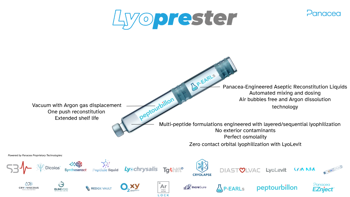
That is the class of problem this company builds instruments for: not the molecule, but the physical and chemical environment that decides whether a molecule survives being made into a medicine. The Lyoprester™ dual-chamber cartridge holds a dried solid and its reconstitution liquid apart until the moment of use — which, for a molecule whose native biology is store inert, activate on arrival, is not packaging around the mechanism but a continuation of it. TgShift™, Cryolapse™ and LyoLevit™ govern the drying that gets it into that cartridge intact.
Follow a synthesised peptide forward and there are five places to lose it — purity, adsorption, degradation, physical state, container — and four of them fall after the chemistry is finished. Nearly everyone supplying peptides owns the first. Panacea Bio Chem owns all five, because it built the instruments for the other four itself, in-house, with the process parameters held internally. Set against a supply chain that stops at synthesis and hands the remaining four steps to somebody else, that is why the peptides leaving this programme are the finest obtainable anywhere. It is a conclusion the chain forces rather than a claim laid over it.
Panacea peptides — Peptourbillon™ dual-chamber presentations ↗
8. Application fields — where this reaches furthest
Because biofilm sits underneath so many persistent problems, the ideas here reach well past a single peptide. Directions worth watching, offered as directions rather than as results:
None of these is an approved use of anything described on this page.
Who publishes this

“Panacea Bio Chem is not merely interested in molecules. Panacea works on the physical, chemical and technological environment surrounding them.”
Bogdan Dicoias
Biochemist and inventor · Director of Panacea Bio Chem
Inventor of the Lyoprester dual-chamber cartridge architecture and of the Cryolapse, TgShift, LyoLevit, DiastolVAC, OxyDeplete and S3Pulse technologies. Panacea Bio Chem →
 Panacea IQP
Frequently asked
What is a biofilm?
A community of bacteria attached to a surface and enclosed in a matrix they secrete themselves — polysaccharide, protein and extracellular DNA. It is a different physical and physiological state from the same bacteria floating freely, and it is that state, not the genome, which resists treatment.
Why do antibiotics fail against biofilm?
Three reasons compound: the matrix slows the drug reaching the deepest cells; those cells are often slow-growing or dormant, and most antibiotics attack an active process; and the structure impedes immune clearance too, so drug and body are blunted together. The concentration needed can be orders of magnitude higher than for the same organism in suspension.
How do you break down biofilm?
Where physical removal is possible it is the surest route — debridement, or taking out an infected device. Chemically, the research interest is in agents acting on the matrix and the membrane rather than on a growth process, which is why cationic peptides are studied here. Nothing described on this page is an approved treatment for anything.
What is LL-37, and where does it come from?
A 37-residue peptide with a net charge of about +6, cut from a larger precursor called hCAP18 — the only cathelicidin humans make — by proteinase 3, after neutrophils release it. The precursor itself is inert.
Does LL-37 break down biofilm?
Published work reports interference with biofilm below growth-inhibitory concentrations, and synergy with polymyxin B against biofilm cells of E. coli and P. aeruginosa. Read those as findings in specific model systems rather than a general property, and note there is no approval anywhere.
Is vitamin D really connected to the body's antibiotics?
Yes, and directly. The CAMP gene is a direct transcriptional target of the vitamin D receptor, and 1,25-dihydroxyvitamin D3 strongly upregulates cathelicidin expression in myeloid cells. That sets how much precursor exists — a separate question from whether it gets activated.
What is the difference between hCAP18 and LL-37?
hCAP18 is the whole 18 kDa precursor and is inert; LL-37 is the active 37-residue peptide cut from its C-terminus. The distinction is not academic — in Papillon-Lefèvre syndrome the precursor is present, the cut cannot happen, and the clinical consequence is severe. There is a page on exactly this.
Is any of this an approved treatment?
No. LL-37 has no marketing authorisation in any jurisdiction. The randomised work that exists is in hard-to-heal venous leg ulcers and is investigational. Nothing here is medical advice.
Trending in the field
Recent developments in the field — refreshed 2026-09-09 by Panacea Bio Chem.
- Coordination chemistry and antimicrobial activity of LL-37, its C-terminal fragment, and a peptidomimetic analogue — PubMed, 2026 Sep 8
- Emerging antimicrobial peptides in gastrointestinal disorders: Dual role in immunity and therapy — PubMed, 2026 Sep 5
- Plasma levels of antimicrobial peptide LL-37 as a biomarker of sleep quality in patients with obstructive sleep apnea: A clinical observational study — PubMed, 2026
- Targeted delivery of antimicrobial peptide LL-37 via ferritin fusion improves antibacterial and anti-inflammatory outcomes in a murine sepsis model — PubMed, 2026 Aug 31
- Combined FMEA and Cause-Effect Analysis Method for Risk Assessment of Failures Impacting Hemodialysis Water Quality — PubMed, 2026 Sep 7
Weekly review — 7–13 Sep 2026
Publications indexed in PubMed in the last 30 days for ("LL-37" OR hCAP18 OR hCAP-18 OR cathelicidin) AND (antimicrobial OR biofilm OR immunomodulat*) — refreshed weekly.
- Building Blocks of 2,3-Diaminopropionic Acid Functionalized with an Oxa-Acid Spacer Shift the Functional Profile of Human Cathelicidin LL-37: Enhanced Enzymatic Stability and Selective Suppression of Neutrophil Degranulation — Pharmaceuticals (Basel, Switzerland), 2026 Jul 24
- Occupational Contact Dermatitis in the Post-COVID Era: From Barrier Dysfunction and Microbiome Dysbiosis to Prevention and Precision Management — Journal of clinical medicine, 2026 Aug 17
- Exploratory Evaluation of Susceptibility Patterns of Clinical Isolates of Acinetobacter baumannii Towards Polymyxins and Different Cationic Antimicrobials — Antibiotics (Basel, Switzerland), 2026 Jul 27
- Combinatorial therapy of LL-37 and ADSCs accelerates diabetic wound repair by orchestrating NRROS-mediated crosstalk between TGF-β/SMAD and hippo/TEAD1 axes — Cytokine, 2026 Aug 25
- Combined FMEA and Cause-Effect Analysis Method for Risk Assessment of Failures Impacting Hemodialysis Water Quality — Therapeutic apheresis and dialysis : official peer-reviewed journal of the International Society for Apheresis, the Japanese Society for Apheresis, the Japanese Society for Dialysis Therapy, 2026 Sep 7
References & further reading
Every source below was retrieved and read at the identifier given. Where a claim rests on a specific study, the number points at that study and not at a review of it.
- Human cathelicidin, hCAP-18, is processed to the antimicrobial peptide LL-37 by extracellular cleavage with proteinase 3Sørensen OE et al. · Blood, 15 June 2001;97(12):3951–9. Identified the enzyme, established that cleavage occurs on exocytosed material rather than intracellularly, and recorded the species difference against elastase-cleaved bovine and porcine cathelicidins.PMID 11389039
- Processing of seminal plasma hCAP-18 to ALL-38 by gastricsin: a novel mechanism of generating antimicrobial peptides in vaginaSørensen OE et al. · J Biol Chem, 1 August 2003;278(31):28540–6. Source for the inactive seminal-plasma form, the pH-dependent gastricsin cleavage, the 38-residue product and its activity equal to LL-37.PMID 12759353
- Kallikrein expression and cathelicidin processing are independently controlled in keratinocytesJ Invest Dermatol, May 2010. The keratinocyte route, and the independence of the two controls.PMID 20090765
- Epidermal proteases in the pathogenesis of rosaceaJ Investig Dermatol Symp Proc, December 2011. Context for dysregulated cathelicidin processing in skin.PMID 22076323
- Human cathelicidin antimicrobial peptide (CAMP) gene is a direct target of the vitamin D receptorFASEB J, July 2005. The primary demonstration that CAMP is a direct VDR target gene, strongly upregulated in myeloid cells by 1,25-dihydroxyvitamin D3.PMID 15985530
- Vitamin D as an inducer of cathelicidin antimicrobial peptide expression: past, present and futureJ Steroid Biochem Mol Biol, July 2010. Review consolidating the vitamin D–cathelicidin literature.PMID 20302931
- Lack of cathelicidin processing in Papillon-Lefèvre syndrome patients reveals essential role of LL-37 in periodontal homeostasisEick S et al. · Orphanet J Rare Dis, 27 September 2014;9:148. Cathepsin C loss-of-function removes the activating step; the precursor is present and the mature peptide is not.PMID 25260376
- Papillon-Lefèvre syndrome patient reveals species-dependent requirements for neutrophil defensesJ Clin Invest, October 2014. A parallel examination of the same processing defect.PMID 25244098
- Biofilm tolerance to antimicrobials — the concentration gapThe order-of-magnitude difference between killing free-floating and biofilm-encased cells of the same organism is a general finding of the biofilm literature rather than a single study, and is described here as commonly cited rather than as a measured constant.PMC10305407
- Synergy between human peptide LL-37 and polymyxin B against planktonic and biofilm cells of Escherichia coli and Pseudomonas aeruginosaAntibiotics (Basel), 15 February 2023. Source for the antibiotic-synergy claim against biofilm cells specifically.PMID 36830299
- Evaluation of LL-37 in healing of hard-to-heal venous leg ulcers: a multicentric prospective randomised studyWound Repair Regen, November 2021. The larger of the two controlled human studies.PMID 34687253
- Treatment with LL-37 in hard-to-heal venous leg ulcers — a randomised, placebo-controlled clinical trialWound Repair Regen, September–October 2014. Title rendered here in neutral wording; the published title uses a term this site does not print in its own copy. Follow the identifier for the paper as indexed.PMID 25041740